

How has technology changed ophthalmic practice?
● Faster, more accurate diagnostics — Automated instruments can collect precise measurements
quickly and consistently.
● Better visualization — Advanced imaging such as OCT allows providers to see detailed cross-sectional
images of structures within the eye.
● Improved disease detection — Modern imaging and diagnostic technology can help identify ocular
conditions earlier and monitor changes more effectively
● Greater patient education — Digital images give providers the ability to show patients what is
happening inside their eyes and better explain their diagnosis
● Increased importance of equipment maintenance — As practices become more dependent on
sophisticated technology, keeping equipment calibrated, maintained, and functioning properly becomes
increasingly important.
https://www.bing.com/images/search?view=detailV2&ccid=GHEyXP%2Fd&id=C826B29758D23EE1B6532707F6D91A0228BCD38E&thid=OIP.
GHEyXP_d5MsSxIapzMm-1gHaDt&mediaurl=https%3A%2F%2Fcanonstreetoptometrists.co.nz%2Fwp
content%2Fuploads%2F2023%2F03%2Foct-blog-header
w1200h600%402x.jpg&cdnurl=https%3A%2F%2Fth.bing.com%2Fth%2Fid%2FR.1871325cffdde4cb12c486a9ccc9bed6%3Frik%3DjtO8KAIa2fY
HJw%26pid%3DImgRaw%26r%3D0&exph=1200&expw=2400&q=tomography++oct&FORM=IRPRST&ck=620BE5093C0F41619D90C96FAF1DF
146&selectedIndex=43&itb=0&cw=1500&ch=860&ajaxhist=0&ajaxserp=0
Ophthalmology practice modernization
Walk into an ophthalmology practice today and you’ll see technology that would have seemed almost
unimaginable to earlier generations of eye-care professionals.
Digital imaging. Automated measurements. High-resolution retinal scans. Electronic records. Advanced
diagnostic equipment. Connected systems that allow physicians to capture, store, compare, and analyze
information in ways that simply weren’t possible decades ago.
But getting here didn’t happen overnight.
The modern ophthalmology practice is the result of more than a century of innovation—and every
generation of equipment has helped move eye care forward.
The Earlier Days of Ophthalmology
For much of the Evolution of Diagnostic Equipment in Ophthalmic Practice, physicians relied heavily on
their own eyes, experience, and relatively simple optical instruments.
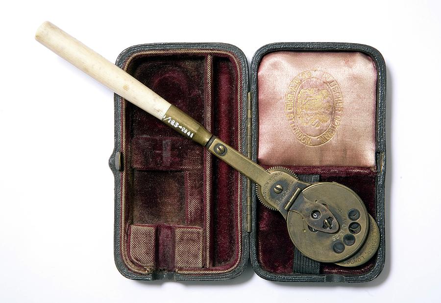
The development of the direct ophthalmoscope in 1851 was a major breakthrough because it allowed
physicians to examine structures inside the eye in an entirely new way. Decades later, the slit lamp
became another defining advancement. Allvar Gullstrand developed the modern predecessor of the slit
lamp in 1911, creating a way for clinicians to examine the eye under magnification and focused
illumination. (EyeWiki)
These instruments became foundational to ophthalmic care.
And many of the instruments that became standard decades ago are still recognizable today.
The difference?
The technology surrounding them has changed dramatically.
From Manual Measurements to Automated Precision
Older practices depended much more heavily on manual measurements and adjustments.
Today, automated refractors, computerized diagnostic instruments, digital tonometry, advanced visual
field systems, retinal cameras, and other technologies can collect measurements quickly and
consistently.
Automated refractors, for example, can estimate a patient’s refractive error before the provider fine
tunes the prescription. (EyeWiki)
That doesn’t eliminate the expertise of the ophthalmologist or optometrist—it gives them better
information to work with.
The goal has always been the same:
better information leads to better clinical decisions.
From Film to Digital Imaging
One of the most noticeable changes is how ophthalmic images are captured and stored.
Historically, retinal imaging relied on film and could involve tedious processes for capturing and
managing images. Digital imaging transformed that workflow, making it possible to capture, store,
review, and compare images much more efficiently. (EyeWiki)
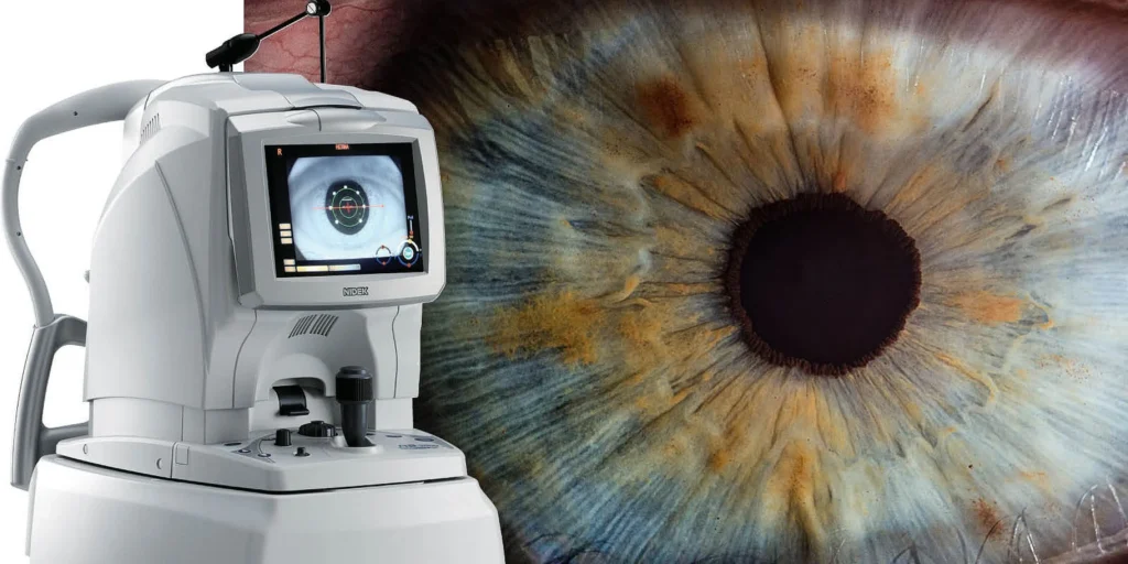
Then came Optical Coherence Tomography, or OCT for short.
Introduced into ophthalmic practice in the 1990s, OCT changed the way clinicians could visualize the
retina by providing high-resolution cross-sectional images of ocular structures. Modern OCT technology
can provide detailed information that helps physicians diagnose disease and monitor changes over time.
(EyeWiki)
In other words, ophthalmology moved from simply looking at the eye to being able to see layers and
structures within it in remarkable detail.
The Modern Ophthalmology Practice
Today’s practice can look less like a collection of individual instruments and more like an interconnected
technology environment.
A patient may move through several diagnostic stations in a single visit. Measurements are captured
digitally, images are stored electronically, and physicians can compare current findings with previous
examinations.
Electronic health records and digital imaging have also created opportunities to bring clinical
information and imaging together in ways that weren’t possible in earlier practices. (National Eye
Institute)
And the evolution isn’t stopping.
Researchers are already working on increasingly sophisticated imaging technologies and artificial
intelligence applications. The National Eye Institute has highlighted advances that combine AI with
ocular imaging to improve resolution and potentially bring cellular-level imaging closer to everyday
clinical use. (National Eye Institute)
Old Equipment Doesn’t Mean Outdated Equipment
One important thing can sometimes get lost in the conversation about modernization:
Newer isn’t always the same thing as better.
Many established ophthalmology practices continue to rely on equipment that has provided years of
dependable service. A well-maintained slit lamp, phoropter, tonometer, microscope, or other
ophthalmic instrument can remain an important part of a practice’s workflow.
Modernizing a practice doesn’t necessarily mean replacing everything.
Sometimes it means maintaining what works, upgrading what makes sense, integrating newer
technology where it provides value, and making sure every piece of equipment performs the way it
should.
That’s where professional ophthalmic service becomes especially important.
The Equipment Behind the Patient Experience
Patients may notice the physician, the staff, and the technology in front of them—but they don’t always
see everything happening behind the scenes.
Every diagnostic instrument needs proper maintenance.
Every piece of equipment needs to be evaluated for performance.
And when something isn’t functioning properly, the impact can extend beyond the equipment itself. It
can affect workflow, productivity, patient experience, and the practice’s ability to provide care
efficiently.
As ophthalmology continues to advance, the need for knowledgeable equipment service doesn’t
disappear. It becomes even more important.
Looking Ahead
The story of ophthalmology is really a story of continuous improvement.
From the earliest optical instruments to the modern slit lamp.
From manual measurements to automated diagnostics.
From film photography to digital retinal imaging.
From simply viewing the retina to generating detailed cross-sectional images with OCT.
And now, toward increasingly sophisticated imaging, automation, connectivity, and artificial intelligence.
The technology may continue to change—but one thing remains constant:
The quality of the equipment matters.
At Sunshine Ophthalmic, we believe that keeping ophthalmic equipment performing at its best is an
essential part of keeping practices running at their best.
Because the future of eye care isn’t just about having the newest technology.
It’s about having the right technology, properly maintained, properly serviced, and ready to perform
when your practice needs it.
The future of ophthalmology is already here. Let’s keep your practice ready for what’s next.
Sunshine Ophthalmic
Ophthalmic Equipment Service & Support
877-801-9503
customerservice@sunoph.com
www.sunoph.com